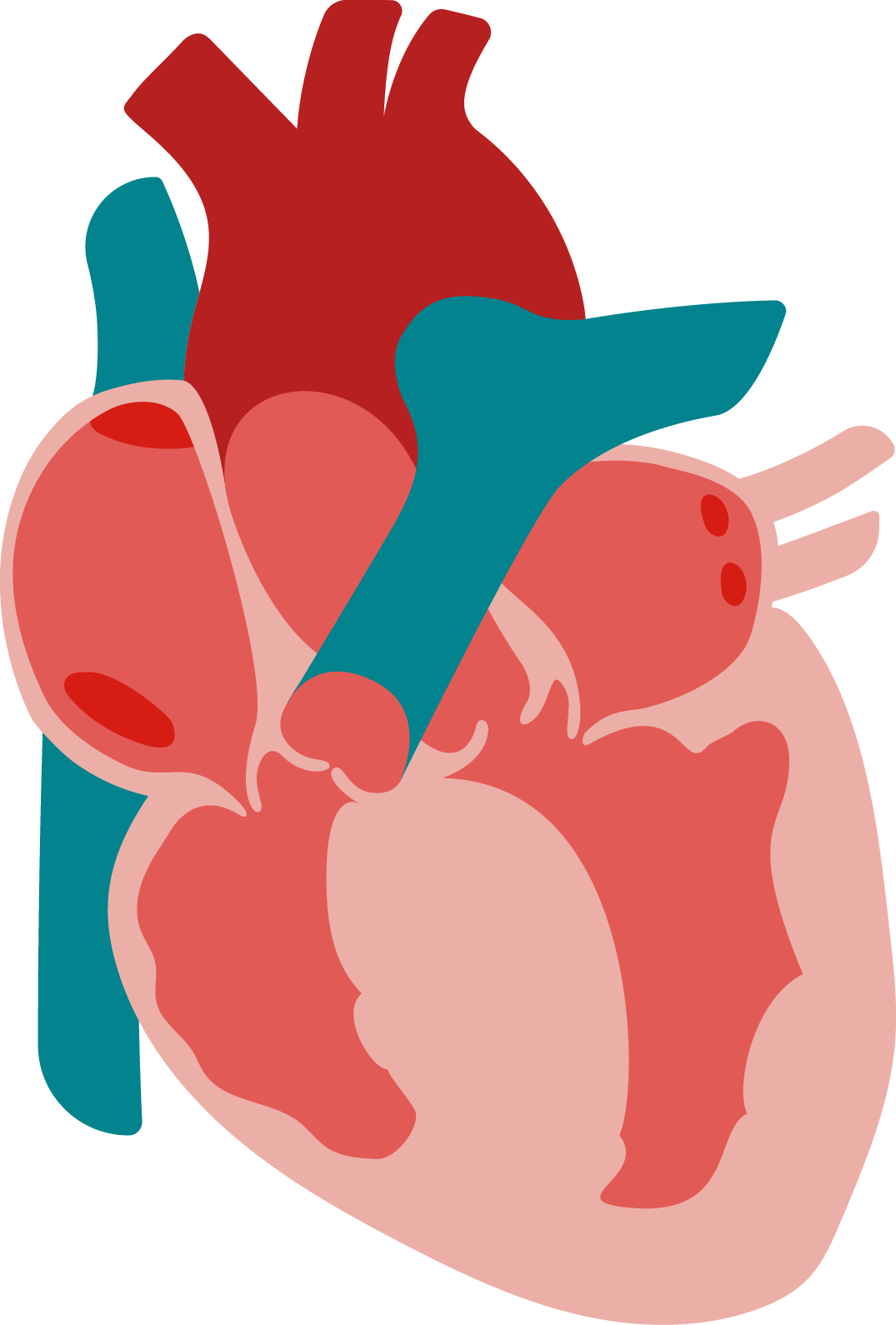
Ventricular septum
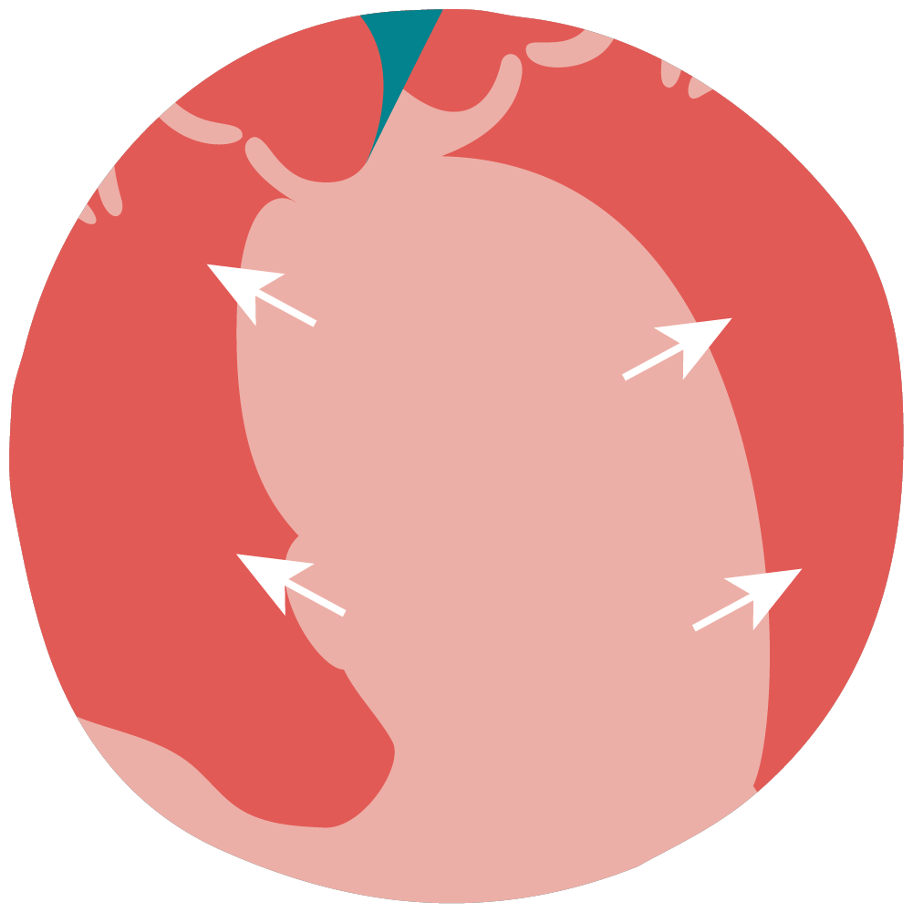
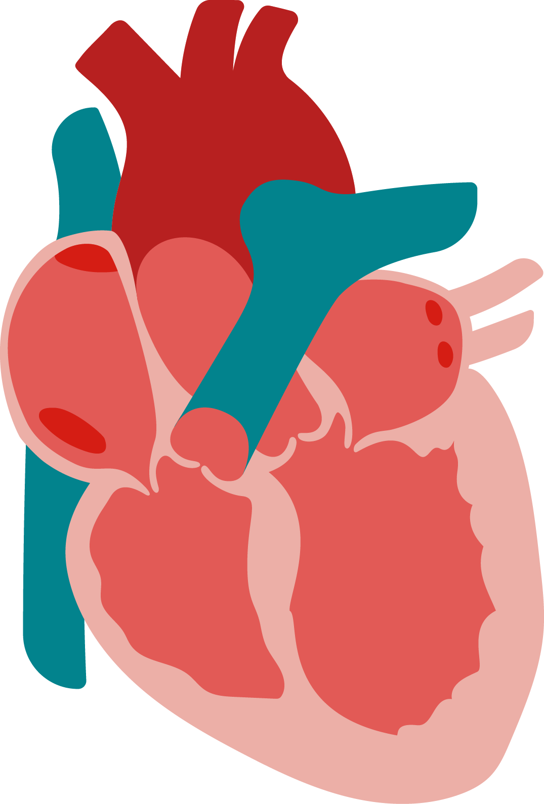
This is the section between the two ventricles (lower chambers) of the heart. In HCM, the muscles in the ventricular septum become thicker, making this part of the heart larger. This affects the function and volume of the ventricles, especially the left ventricle. A thicker ventricular septum also makes the muscle less flexible. This can make it harder for your heart to pump blood.
What Does Hypertrophic
Cardiomyopathy Look Like In the Body?
End Tour


healthy heart
Hypertrophic Cardiomyopathy
healthy heart
Hypertrophic Cardiomyopathy


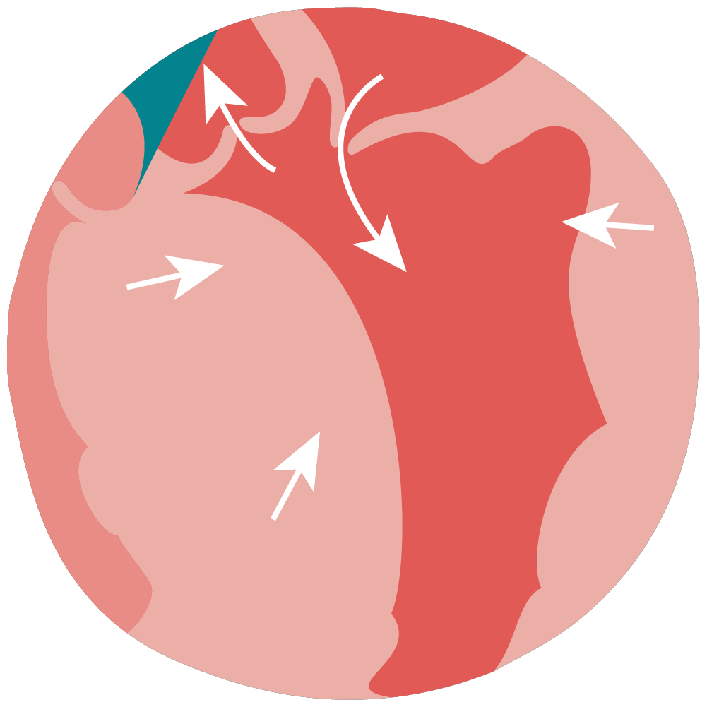
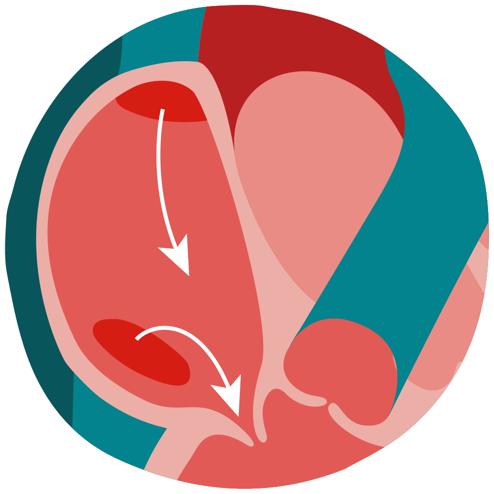
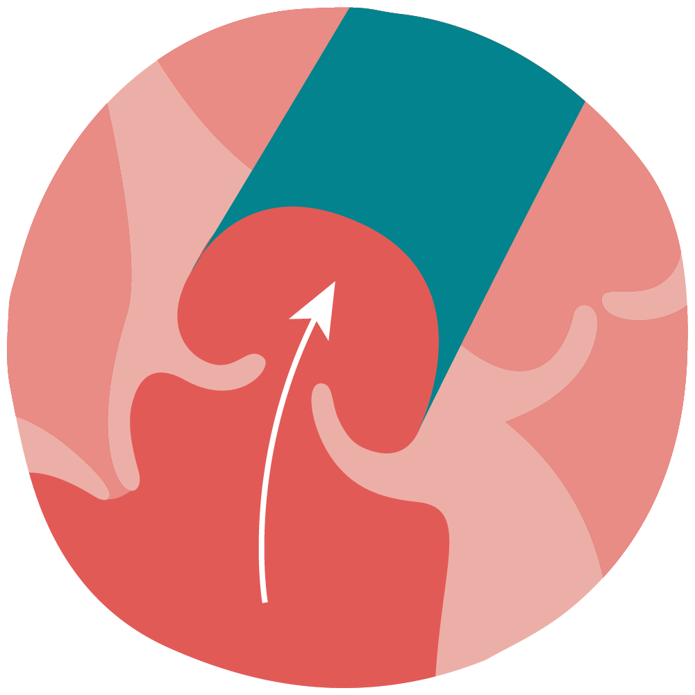
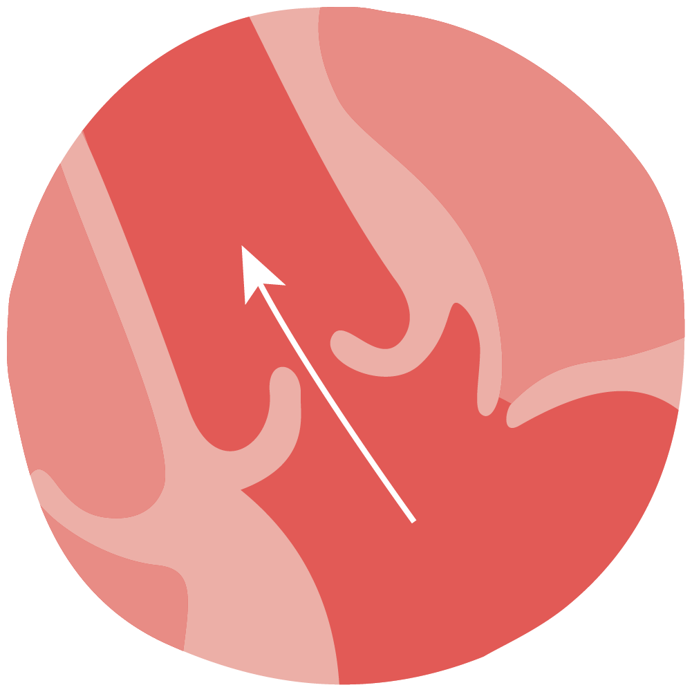
In HCM, the ventricular septum takes up more space, shrinking the size of the left ventricle. Oxygen-rich blood enters the left ventricle before getting pumped out to the rest of the body. HCM can make it harder for the left ventricle to take in and pump the amount of blood needed to meet the body’s needs.
Left ventricle
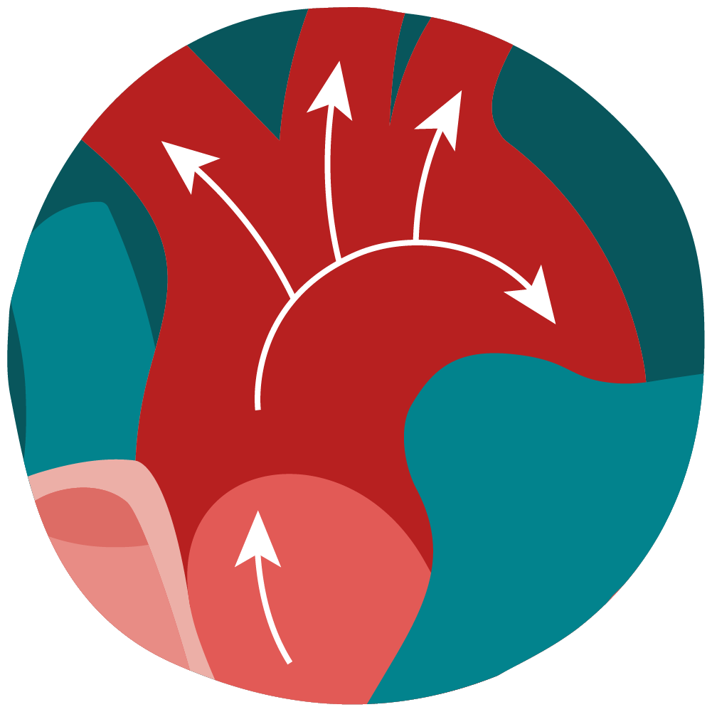
Oxygen-rich blood is pumped from the left ventricle to the aorta, where it is sent around the body. A thicker ventricular septum can block or reduce this normal blood flow. Many of the symptoms of HCM are due to changes in blood flow.
Blood flow


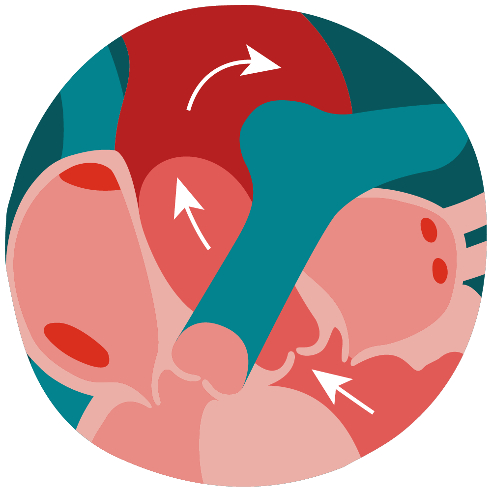
While HCM predominantly affects the left ventricle, in later stages of the disease, the right ventricle can be affected. The right ventricle sends low-oxygen blood to the lungs to get more oxygen. Blood travels to the lungs through the pulmonary arteries. When the right ventricle is involved in HCM, it is not able to efficiently pump blood to the lungs, which can lead to shortness of breath and swelling.
Right ventricle
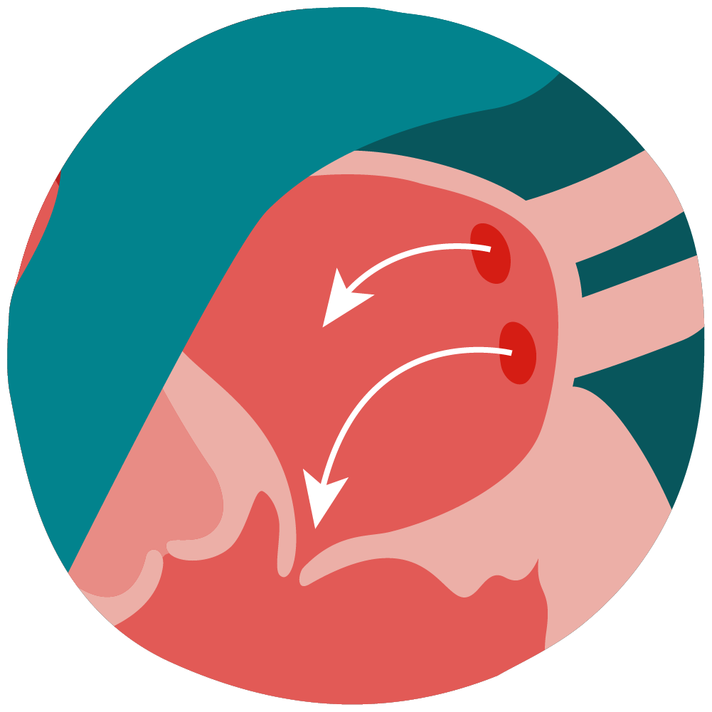
The right atrium gathers low-oxygen blood from around the body. When the right atrium fills with blood, the tricuspid valve opens to let blood into the right ventricle. From there, it travels to the lungs to get more oxygen.
Right atrium
The tricuspid valve regulates blood flow from the right atrium to the right ventricle. The right atrium fills with blood that is low in oxygen and high in carbon dioxide. Once the atrium fills, the tricuspid valve opens to allow blood to fill the right ventricle.
Tricuspid valve

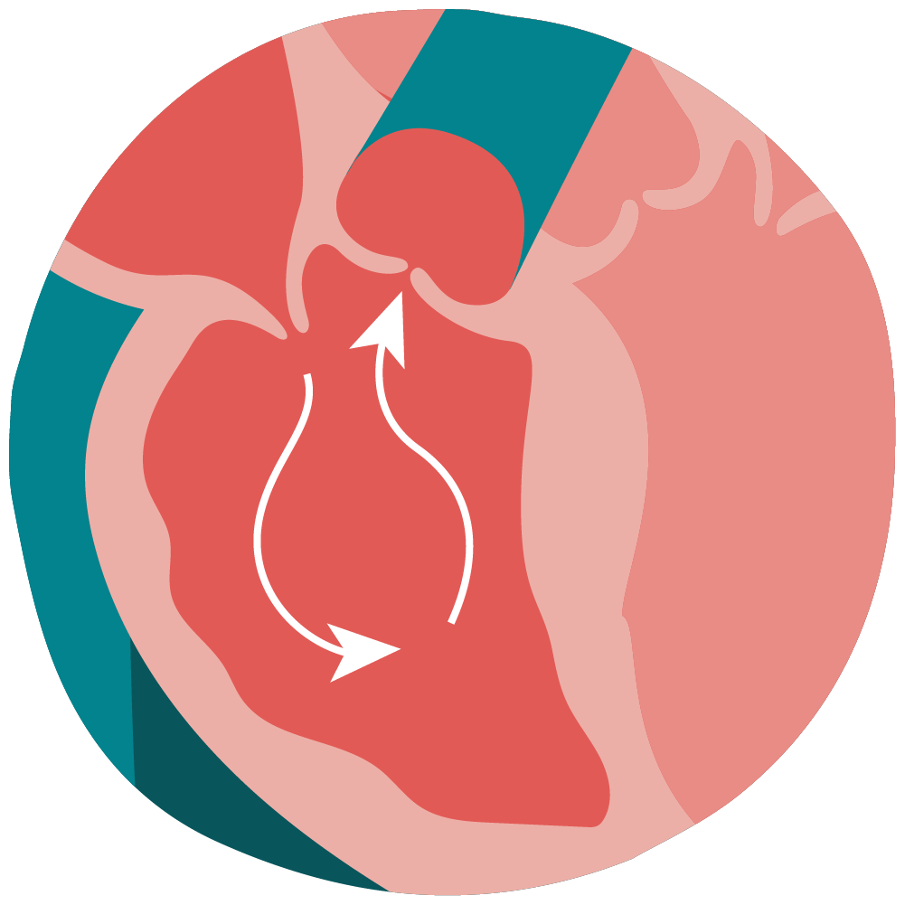
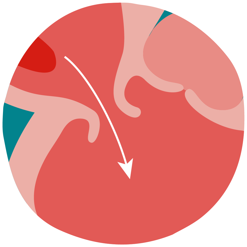
Blood travels from the left atrium into the left ventricle through the mitral valve. HCM changes
the shape and blood flow of the left ventricle. Sometimes, the mitral valve is affected by HCM, causing blood to flow backward from the left ventricle into the left atrium. This is known as mitral valve regurgitation.
Mitral valve

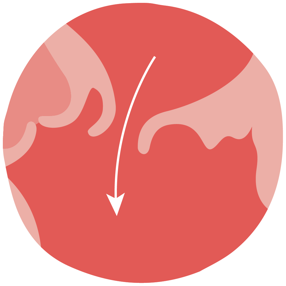
This valve controls blood flow between the right ventricle and the pulmonary artery. Blood from the right ventricle is low in oxygen and high in carbon dioxide. The pulmonary artery sends blood to the lungs. The lungs exchange the carbon dioxide gas in the blood for more oxygen.
Pulmonary valve

This valve is between the left ventricle and the aorta. It opens and closes to control the flow of oxygen-rich blood from the left ventricle into the aorta. The aorta is connected to a network of arteries to supply the body with oxygen and nutrients.
Aortic valve

The aorta is the largest artery in your body. Oxygen-rich blood is pumped from the left ventricle into the aorta. From there, it branches out into smaller arteries to supply blood throughout the body. The thicker wall of the left ventricle can reduce the amount of blood that gets pumped to the aorta. This means that the body may not be able to get enough blood and oxygen.
Aorta

The left atrium is the first stop for blood after it gets an oxygen boost from the lungs. Blood gathers here and then enters the left ventricle through the mitral valve. HCM can reduce how much blood is able to flow into the left ventricle. Blood may backflow into the left atrium if there is no space for it to pass into the left ventricle, which can lead to enlargement of the left atrium.
Left atrium


